Extra 8% Off sitewide with "PD8"
Free shipping


Add optional protection for eligible accidental damage and service after the original warranty.
Optional. Added as a separate cart item. Terms, conditions and exclusions apply.
We provide free home delivery for orders over $30.
We ship from our CA warehouse whenever stock is available, with delivery typically taking 3–7 days. If items are dispatched from our international warehouse, delivery may take 7–15 days.
All orders are trackable, and you will receive an email with tracking details once your order has been shipped.
With our clinically tested and proven system, we are confident in what we deliver.
You may request a full refund by returning your undamaged products within 30 days. Once the item is received at the Freyara warehouse and passes inspection, the refund process will begin.
For detailed information regarding refunds, please contact our customer service at suri@freyara.com
Freyara offers a one-year warranty to ensure your shopping experience is worry-free.
If your product experiences any quality issues within one year of purchase, simply provide your order receipt and contact our customer service team at suri@freyara.com .
We will assist you with confirmation and provide safe, convenient repair or replacement services.
We'll always be there for you.
We provide product tutorials and dedicated support just for you!
If you have any questions, please feel free to contact our customer service team at suri@freyara.com
.
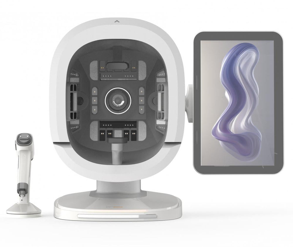
* Comprehensive 180° Skin Analysis with 8 Spectral Imaging Modes Capture your client’s entire face in one click – front, left, and right simultaneously – for a complete blind‑spot‑free view. Eight specialized light sources (White, Polarized, Wood’s, UV, Brown, Red, Mixed) reveal surface blemishes, deep pigmentation, inflammation, collagen loss, and more, providing dermatologist‑level insights invisible to the naked eye.
* AI-Powered 30+ Detection Dimensions + Customized Skin Color Algorithms Go beyond basic analysis with 30+ professional indicators: moisture, pores, wrinkles, acne, pigmentation, sensitivity, skin age, pH, and collagen. Industry‑first skin‑color optimization adapts algorithms for yellow, white, black, and brown skin tones, ensuring accurate results for every client.
* Instant Report Generation & 16-Type Baumann Classification Generate a comprehensive, customizable report in seconds with one‑click operation. Includes the internationally recognized 16‑type Baumann skin classification, pH value, skin properties (dry/oily/combination/normal/sensitive), and color‑coded grading (A–E). Clients can access their reports anytime via QR code on mobile.
* Advanced Microscopic Imaging & 3D Layer Analysis The handheld micro‑imager with white, polarized, and UV light lets you inspect skin and scalp details at both surface and deep levels. Use the 3D slicing function to visualize epidermis and dermis layers, and the muscle‑bottom perspective to compare pigmentation, redness, and inflammation between superficial and basal layers.
* Professional Data Management & User‑Friendly Operation Designed for clinics, spas, and retailers: one‑click capture, image export, backend data management, and parameter adjustment for fine‑tuning results. Supported by patents and certifications (CE, ROHS). Ideal for consultation, treatment planning, and tracking progress – all in a sleek, ergonomic device.


| Feature | Benefit |
|---|---|
| 180° Blind-Spot-Free Capture | Three fixed perspectives (front, left, right) for complete facial analysis |
| 8 Spectral Imaging Technologies | Reveal skin conditions invisible to the naked eye |
| AI-Powered Analysis | 30+ detection dimensions with professional-grade accuracy |
| One-Click Operation | Instant photo capture and report generation |
| Multi-Skin Tone Optimization | Industry-first customized detection for all skin colors |
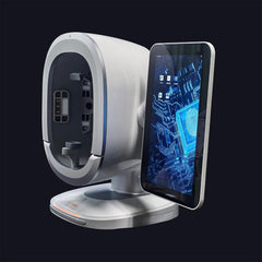
The K33 AI Intelligent Skin Tester represents a comprehensive upgrade in skin analysis technology. Designed for dermatologists, aesthetic clinics, and premium skincare retailers, it delivers professional, objective analysis of facial skin conditions with unprecedented accuracy and efficiency.
| Mode | What It Reveals |
|---|---|
| White Light | Surface blemishes, acne, spots, wrinkles, pores under natural light |
| Positive Polarized Light | Skin texture, fine lines, wrinkles, bumps, acne scars |
| Negative Polarized Light | Surface irregularities and texture analysis |
| Wood's Light | Deep dermal pigmentation with enhanced contrast |
| UV Light | Bilirubin distribution, pigmentary disorders, infections |
| Brown Light | Subcutaneous melanin accumulation, UV damage assessment |
| Red Light | Hemoglobin distribution, inflammation, sensitivity, redness |
| Mixed Light | Collagen loss evaluation and skin roughness analysis |
| Skin Tone | Optimized Algorithm |
|---|---|
| Yellow Skin | ✓ |
| White Skin | ✓ |
| Black Skin | ✓ |
| Brown Skin | ✓ |
The K33 adapts its detection algorithms and evaluation dimensions based on the unique characteristics of different races and skin tones. The 3D model display automatically adjusts according to selected skin color.
| Category | Indicators |
|---|---|
| Hydration & Barrier | Moisture, Skin Barrier, Sebum |
| Pores & Texture | Pores, Blackheads, Roughness |
| Pigmentation | Superficial Pigment, Deep Pigment, Brown Pigment, Sunspots, Mixed Spots |
| Aging Signs | Wrinkles, Collagen, Skin Age |
| Sensitivity | Heat Map of Sensitivity, Redness Map |
| Skin Conditions | Acne, Porphyrin |
| Tool | Function |
|---|---|
| 3D Panoramic View | Front face, left cheek, and right cheek perspectives |
| Muscle Bottom Perspective | Compare superficial and basal layer issues (pigmentation, redness, inflammation) |
| 3D Slicing | Layered scanning of epidermis and superficial dermis |
| Microscopic Detection | Handheld micro-imager for skin and scalp details |
| Annotation Pen | Mark visible issues directly on images for consultation |
Best For
The K33 is designed for professional skin consultations that benefit from complete three-angle facial capture, clear report sharing and optional close-up skin or scalp inspection.
Prepare the Workstation
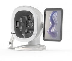
Place the analyzer on a stable, dry surface with consistent room lighting. Confirm the 15.6-inch display, three-camera imaging unit and 50× handheld microscope are connected and clean.
Prepare the Client
Remove makeup, sunscreen and excess surface oil; keep hair away from the face; create or open the correct client profile; and select the appropriate skin-tone algorithm.
Capture and Explain
Position the client once for simultaneous front, left and right imaging, run the eight spectral modes, review the relevant 30+ indicators and use the microscope for localized detail. Generate the report, review it with the client and provide the QR code when mobile access is desired.
Track Progress
Repeat later sessions with similar preparation, lighting and positioning so date-to-date comparisons remain useful.
White Light — Reviews visible surface blemishes, acne, spots, wrinkles and pores under natural-looking illumination.
Positive Polarized Light — Enhances fine lines, texture, pores, bumps and acne-scar patterns.
Negative Polarized Light — Reduces surface reflection to support assessment of pigmentation, capillaries and skin uniformity.
Wood's Light — Adds contrast for deeper-looking pigmentation.
UV Light — Highlights fluorescence, porphyrins and selected pigment- or pore-related conditions.
Brown Light — Supports visualization of UV-related damage and subcutaneous melanin patterns.
Red Light — Emphasizes hemoglobin distribution, redness, inflammation and sensitive-looking areas.
Mixed Light — Supports collagen-loss and surface-roughness analysis.
50× Microscope — Uses white, polarized and UV imaging for localized skin or scalp inspection.
3D Tools — Panoramic view shows the facial angles together; slicing and muscle-bottom views help compare surface and deeper-looking layers.
Report and QR Modes — Use the full report for consultation, save results for later comparison and share client-facing access by QR code.
1. Power on the K33 and verify the three-camera system, display and 50× microscope.
2. Clean the client's face and remove makeup, sunscreen and excess oil.
3. Create or open the correct profile and select the skin-tone algorithm.
4. Position the client according to the on-screen guide for simultaneous front, left and right capture.
5. Run all eight spectral modes without changing the client position or room lighting.
6. Review the relevant moisture, sebum, pores, wrinkles, acne, pigmentation, sensitivity, pH, skin-age and collagen indicators.
7. Use the 3D views to explain surface and deeper-looking patterns.
8. Use the handheld microscope only when a selected skin or scalp area needs closer inspection.
9. Review the Baumann skin type and generated recommendations with the client.
10. Save the session, generate the QR report and clean all client-contact surfaces before the next use.
Brand: FREYARA
Model: K33
SKU: FY2201.0001.0030
Product Type: Professional AI Skin Analysis System
Display: 15.6 Inches
Facial Capture: 180° Front / Left / Right Simultaneous Imaging
Spectral Imaging Modes: 8
Detection Dimensions: 30+
Skin-Tone Algorithms: Yellow / White / Black / Brown
Skin Classification: Baumann 16 Types
Localized Imaging: 50× Handheld Skin and Scalp Microscope
Analysis Views: 3D Panoramic / 3D Slicing / Muscle-Bottom Perspective
Report Delivery: On-Screen and QR-Code Mobile Access
Certifications Listed: CE and RoHS
This equipment is covered by FREYARA's one-year limited warranty. For an operating or hardware issue, contact customer support with the order number, a clear description and photos or video. Support may include guided troubleshooting and covered repair or replacement under the published warranty terms. Damage caused by misuse, unauthorized modification, improper storage, liquid exposure or failure to follow the supplied instructions is not covered. Returns remain subject to the published 30-day return conditions.
For professional cosmetic imaging and consultation use. The K33 does not diagnose disease and does not replace medical or dermatological assessment. Obtain permission before capturing, storing or sharing facial and scalp images or QR-code reports. Select the correct client profile and skin-tone setting before analysis. Clean the microscope and other client-contact surfaces between users according to the manual. Do not image broken, bleeding or actively infected areas. Operate on a stable, dry workstation with undamaged cables and protect stored personal data in accordance with applicable privacy requirements.
Q: Why does the K33 use three cameras?
A: The three-camera system captures the front, left and right sides of the face at the same time, helping the consultation cover more of the facial surface without separate repositioning.
Q: What can the K33 analyze?
A: It reviews more than 30 displayed indicators, including moisture, sebum, pores, wrinkles, acne, pigmentation, sensitivity, pH, skin age, collagen and other skin-quality measurements.
Q: How does the K33 support different skin tones?
A: The system includes dedicated detection algorithms for yellow, white, black and brown skin tones and also organizes results using the Baumann 16-type system.
Q: What is the 50× microscope used for?
A: It provides close-up white, polarized or UV imaging of a selected skin or scalp area when the main facial scan needs additional localized detail.
Q: Can a client view the report on a phone?
A: Yes. The K33 can generate a QR code that opens the client-facing report on a mobile device, while saved sessions support later progress comparisons.
Q: Is the K33 a medical diagnostic device?
A: No. It is a professional cosmetic skin-imaging and consultation system. Concerning or persistent findings should be referred to a qualified healthcare professional.
Thanks for subscribing!
This email has been registered!